
 English
Κανένα προϊόν στο καλάθι σας.
Εταιρικά Νέα
A Fully Integrated Diagnostic Process – Through Advances in Scanning Technology
by Tim Nolting, Dr MSc, Frédéric Poirier, DDS, and Thomas Giblin, BSc, BDent(Hons)
Abstract
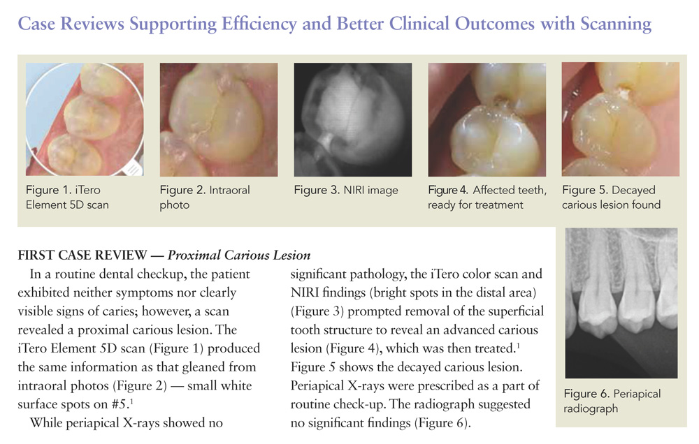
The iTero Element 5D imaging system is the first intraoral 3D scanner integrated with near-infrared imaging (NIRI) technology. NIRI has the potential to revolutionize patient treatment and the overall workflow in dental offices. This technology provides practitioners with an aid for early detection of interproximal caries above the gingiva, which is one of the gravest threats to oral health (equal in seriousness to periodontal disease) per the World Health Organization (WHO).
In the near-infrared electromagnetic spectrum range of 0.7 to 2.0 µm, the iTero Element 5D Imaging System uses light of wavelength (= 850 nm), which interacts with the hard tissue to provide additional data of the tooth structure. The dentin will appear bright, with areas of pathology or demineralization appearing as white spots on the display. The iTero Element 5D imaging system, the latest incarnation of NIRI technology, is an “innovative, integrated optical diagnostic aid,” using a class 1 laser, as Keshav stated in the iTero Element 5D Clinical Guide (Near-infrared imaging technology in dentistry — iTero Element 5D). It gives practitioners the ability to view multiple dimensions of data, as well as to virtually manipulate the model for a comprehensive view. It is the logical next step in digital diagnostic technology and is quickly replacing both conventional impressions and first-generation intraoral scanners.
Advanced scanning technology together with artificial intelligence (AI), streamline the treatment and diagnosis process into the future of dentistry.
Keywords
iTero Element 5D imaging system, patient education, near-infrared imaging (NIRI) technology, dental diagnostics, interproximal caries, restorations, technology adoption, office workflow, practice growth, artificial intelligence (AI)
This white paper has been co-written by 3 dentists who have been using the iTero Element 5D for at least 6 months and refers to a survey of 15 dentists practicing in Germany, Italy, United Kingdom, France, Hong Kong, Australia, and Canada.
Introduction: Impact of Technology Adoption for Practice Growth
In this paper, the ways that adoption and integration of new technologies [particularly, NIRI, the iTero Element 5D imaging system, and artificial intelligence (AI)] will overhaul dental office workflow, optimize diagnosis and treatment planning, and improve practice efficiency are highlighted.
Conventional methods of diagnosing dental caries and other oral pathologies rely on visual and tactile methods coupled with radiography (X-ray). These methods can have significant drawbacks based on visibility, accessibility, and subjective judgment, equal in seriousness to periodontal disease.1
First-generation intraoral scanners (IOS) required the application of powder to the teeth for opacification; this could be clumsy and messy for the practitioner or dental assistant, as well as the patient. Moreover, these early intraoral scanners functioned as little more than digital impression systems. Since then, advances in laser technology and scanning speed, as well as enhanced displays featuring in-color 3D models of the dental arches, like the iTero Element 5D imaging system, have broadened the appeal and functionality of IOS technology for use in general dentistry.1
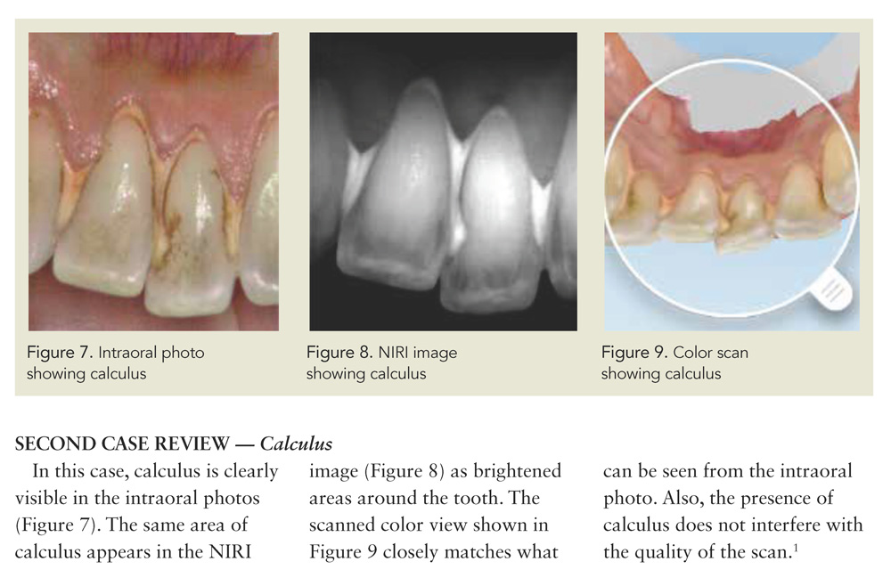
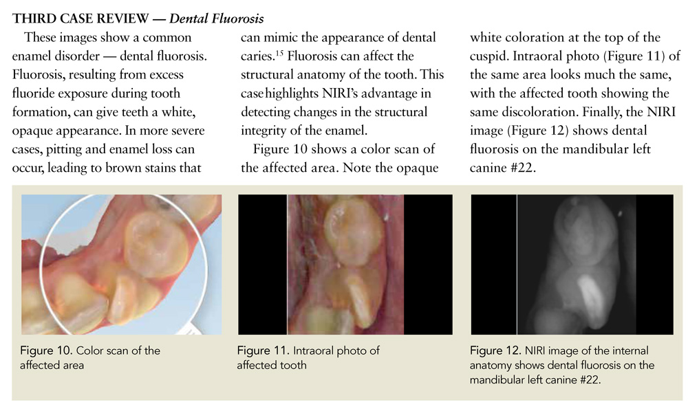
The most cutting edge of these is the use of NIRI for diagnostic imaging, which works by emitting infrared light into the surface of the tooth. The light diffuses through the highly scattering dentin, reflecting off the enamel of the crowns and creating an image of the occlusal surfaces. While much new decay occurs in pits and fissures, and therefore cannot be detected with conventional X-rays because of the overlapping topography of the tooth surface of posterior teeth,2,3 dentists can check for this type of caries with a probe. NIRI scanning is especially useful for detecting interproximal caries above the gingiva that is difficult to see with the naked eye or X-rays, and impossible to detect by probing. In a survey of practitioners who use the iTero Element 5D scanner as part of their diagnostic protocol, 87% of surveyed participants indicated they increased the number of diagnosed interproximal caries above the gingiva by 56% on average. Near-infrared imaging has the potential to allow for superior diagnostic efficiency, particularly when synced with emerging dental AI technologies for enhanced diagnostics and restoration design.
Patient Experience During the Visit
Unlike conventional dental X-rays, NIRI does not expose the patient or the practitioner to ionizing radiation and its potentially harmful effects, and is therefore safe to use whenever a clinician suspects the presence of dental caries or other pathology that may be hidden by enamel.1 A scan can provide more nuanced information and serve as an adjunct to traditional radiographs and intraoral photos, and in some cases even replace conventional diagnostic methods. This a clear advantage, improving patient education and dental office workflow, and reducing risk associated with diagnostic X-rays.
IOS has the broadest indications for clinical use; virtual impressions created with NIRI technology are used in a wide range of procedures in general dentistry and across specialist disciplines, including prosthodontics, implantology, and orthodontics.4 The images can be worked with easily to give a comprehensive view of the oral anatomy. Dental researchers, including those who conducted a 2017 Massachusetts Institute of Technology study of 10 subjects with varying dental conditions, agree that quality of near-infrared images is superior to that of conventional radiographs; they are a better diagnostic aid.3,1,5,6 Likewise, a 2018 study compared NIRI to digital bitewing (DBW) radiography for both intra- and interexaminer reliability, using 12 examiners and 100 images. Reliability on both counts was significantly better with the near-infrared images when used for caries detection.6
Better Patient Communication and Comfort
Patients today are more educated and better informed about their health than ever before. Most want to understand the diagnosis process and be proactive in treatment. However, in a 2013 study on patient understanding and recall by Misra et al., the authors strongly concluded that “patients do not recall as much advice and agreed actions about future dental care as dentists believe they have discussed. These results have implications for patient adherence with oral health instructions.”7
It is reasonable to assume that the disconnect between the information doctors provide and what patients can recall could be improved by utilizing visual aids, including scans. The ability to show patients a picture of their oral health, as opposed to, or as an aid to, merely explaining it to them verbally, is a powerful educational tool with the potential to improve patient compliance. As an example of the power of harnessing technology, a 2018 study of 291 adolescent dental patients showed that the influence of a mobile app for oral health education increased users’ knowledge and produced a measurably better standard of oral hygiene.8 Overall, this indicated that patients respond positively to technological and visual aids.
The iTero Element 5D imaging system has a larger display screen and is built to capture data faster than the previous generations of the Element scanners. These features enable the doctor to evaluate the patient scan chairside and direct a patient’s attention to particular areas shown on the screen as a diagnosis is delivered. As we like to say, a picture is worth a thousand words, and indeed, patients show more confidence and greater understanding in interpreting scanned images alongside their doctors than they do when being shown a dental radiograph. Images produced by the iTero Element 5D imaging system look familiar to the layperson; they closely resemble digital photos and other common computer images that have become ubiquitous in everyday life. This can be helpful in the education of patients and help them to better understand treatment. In fact, out of practitioners surveyed, 100% of users agree that the iTero Element 5D scanner enables better patient education and understanding of their oral health. This, in turn, can translate into increased patient acceptance of treatment. For instance, the same survey found agreement among users that the imaging and visualization capabilities of iTero Element 5D scanner lead to increased patient acceptance of recommended caries treatment.
Patient experience is also augmented due to the fact that the process of taking the scan is often more comfortable than traditional impressions and radiographs. The speed and ability of discussing their images chairside with their doctor also please the patient. Engaging them in this process encourages them to ask questions, thereby allowing the dentist to address any concerns. This ultimately empowers the patients to make well-informed decisions on treatment.
In particular, the time lapse feature distinctly highlights any change over time, whether the topic of concern is tooth wear or movement. The outcome simulator gives a 60-second demonstration of the potential outcome, along with time lapse, which compares scans over time to infer progress.3 Patients can therefore see and easily understand the changes occurring in their mouth. They are much more likely to proceed with treatment when they fully comprehend the situation and the implications of choosing not to treat. With a scan, they can fully visualize what is going on.
Time saved by using an advanced scanning diagnostic aid such as the iTero Element 5D imaging system allows doctors and technicians to dedicate attention to patients’ personal experience and increases their acceptance of recommended treatment. The presence of cutting-edge technology in the dental office fosters patient confidence, as they can see that their doctor uses the most up-to-date diagnostics. This added confidence can further lead to increased acceptance of treatment.
For example, a survey of practitioners who incorporated the iTero Element 5D scanner into their diagnostic protocol found that 79% of participants reported an average increase in patient acceptance of interproximal caries treatment by 71%. In the final analysis, more advanced diagnostics fosters better communication and happier, healthier patients. The combination of patient satisfaction and higher rates of recommended treatment acceptance due to better diagnostics, along with the timesaving efficiency of NIRI scanning, is an equation for boosting practice incomes.
Increased Restorative Cases with Better Clinical Outcomes
The iTero Element 5D imaging system’s overall efficiency creates a more streamlined workflow in the dental office. With the iTero Element 5D, a scan is taken at the beginning of every visit. Other diagnostic methods may or may not be necessary, as the scan does not replace the physical intraoral or extraoral examination. However, it is our experience that an initial scan often eliminates the need for cumbersome, time-consuming X-rays, which would also mean that patients are not subjected to the emission of ionizing radiation.
In his practice, Dr. Nolting found that by using the iTero Element 5D imaging system, approximately 5% more caries was detected than with conventional diagnostics. This is partly attributable to the streamlining effect on office workflow — now doctors using advanced scanners can see more patients because of the reduced time involved, but they can also detect pathologies that might previously have been overlooked. Compared to conventional radiographs, a 3D scan provides a more comprehensive approach that enables the doctor to view all surfaces of every tooth. Thus, scanning is more efficient for revealing interproximal caries decay above the gingiva.
In a survey of practitioners incorporating the iTero Element 5D scanner into their current diagnostic protocol, 79% of survey participants reported an average increase of 32% in the number of treated restorative cases, while reporting an average increase of 57% in the number of treated interproximal caries. These increases resulted in an average hike in business revenue of 25% and 34% for the practice, respectively. Also, in treatment, being able to see into the tooth’s internal anatomy allows dentists to be more conservative with the tooth structure, based on the quality of enamel that is preserved. This leads to increased patient health, preventative efficacy, well- documented practice volume and growth, as well as improved retention of patients. In a survey of iTero Element 5D scanner users, 93% of those surveyed agreed that with the improved communication capabilities of the iTero Element 5D scanner, they expect to improve practice patients’ retention rate. By starting every appointment with a scan, practitioners will have the upper hand in detecting interproximal caries above the gingiva in its earliest stages, even before it shows up on a bitewing radiograph.
Creating Efficiency for Restorative Workflows and Labs
In the past, many dentists have felt pressured to invest in maintaining in-house laboratories for creating accurate restorations. Now, scanning can replace the time-consuming process of creating a model and then using wax to build the teeth back up in the laboratory, which can take a significant amount of time per tooth.
With the iTero Element 5D imaging system, the dental assistant, hygienist, or the dentist performs the scan and hits “send” — it’s that simple.
Models can be delivered to the office within 2–3 days using a lab workflow or fabricated chairside within 1–2 hours using a 3D printer. This replaces the traditional processes that required having a full-time technician on staff and the additional physical space for a lab. A streamlined practice resulting from adopting new digital technologies will need fewer employees and less space, thus positioning NIRI scanning as the default method of monitoring and diagnostics.
In terms of restorations, for example, a major implication is the time savings that can be achieved per crown. Digital impressions have been shown to be a satisfactory alternative to conventional methods for creating impressions.
A 2013 study by Seelbach et al. concluded that digital impression systems allow the fabrication of fixed prosthetic restorations with similar accuracy to that of conventional impression methods.9 Thus, scanning saves precious office time, enabling dentists to outsource many of the tedious steps associated with restorations, and to focus their own efforts on design and finishing. It is also a useful method of documenting ongoing problems and treatment.
Not only useful for crown and bridge work and diagnostics, scanning can be seamlessly incorporated into everyday practice to help practitioners monitor patient oral health. The iTero Element 5D imaging system is more versatile than older generations of scanners, and it is expressly compatible with Invisalign. With Invisalign’s solid comparability behind the iTero, there is a drive to continue to improve design and functionality, to make it more than just a scanner, but a more comprehensive diagnostic aid.
Ease of Use and Accuracy
The iTero Element 5D imaging system offers a light and sleek scanning wand. It is user- friendly; scanning at a rate of 6,000 frames per second, 20 times faster than the earlier models of the iTero scanner with little to no learning curve.10 This system offers screenshot capability as well as various views including intraoral camera, NIRI, and monochrome. A comprehensive archive of instructional videos is available on iTero’s Support website,11 making it simple and easy for technicians to get questions answered and get quick training on how to use the technology in every diagnostic context. The system’s website (myitero.com) also provides the clinician with the ability to store cases, a feature that affords the practitioner the luxury of reviewing cases at their own discretion.
Scanning is noninvasive. When compared to conventional impressions, the use of an intraoral scanner has the ability to improve the patient experience with regard to comfort, gagging, breathability, tastes, and smells. It is easier, cleaner, safer, and more patient-friendly than other diagnostic aids and methods.
Prevention of Harmful Radiation Associated with Radiographs
The advantages of NIRI imaging over X-rays cannot be overstated. Beside the practical advantages — overall time efficiency, labor (and, thus money)-saving, files that are easy to delete and redo, ease of storing files in digital form, and transfer of images between practitioners via electronic transfer,4 the most obvious desirable outcome is eliminating the risk of irradiation for both patient and practitioner. In 2018, Hwang et al. published a review of 2,158 studies to summarize the results of studies of the association between exposure to dental X-rays and health risk. Although the level of exposure from dental diagnostic X-rays is lower than that of medical radiation, there is an innate risk from radiation exposure.12 Therefore, for certain categories of patients, notably those at low risk of developing caries, and also pregnant women, regular bitewing radiographs are neither indicated nor advisable.13,6 Any diagnostic aid or technology that helps eliminate the need for X-rays marks an advance in treatment approach.
Moreover, NIRI technology is shown to be as effective in detecting interproximal caries above the gingiva as radiography,1 perhaps even better — a University of California School of Dentistry study found that with traditional radiography, interproximal caries above the gingiva are undiagnosed up to 40% of the time.14 For conventional X-rays to reliably detect a carious lesion, there must be a certain amount of decay present. A near-infrared image can help the dentist to detect interproximal caries above the gingiva weeks or months before it is severe enough to show up on a conventional radiograph. Starting every appointment with a scan will reduce the number of X-rays taken, and thus reduce exposure to radiation, while increasing diagnostic accuracy. Even in ambiguous cases, where the doctor feels an X-ray is required to be more confident in diagnosis, an initial scan is always an effective aid to rule out an unnecessary step and increase patient confidence.
Evolution of Dental Office Technology
As has been true in other professions, technological advances are streamlining the dental workplace and
helping reduce health risks to clinicians and patients alike. NIRI technology fits in well with the prevailing mode of comprehensive dentistry, as it is a way for clinicians to include the patient, clearly showing them, with easy-to-understand images, the intricate relationship between good oral health and overall well-being. It seems reasonable to extrapolate that NIRI technology should be a useful aid for underscoring the implications of forgoing treatment.
For practices that were already on the way to digitizing much of the paper workflow and daily management (scheduling, communications, etc.), using digital diagnostics actually speeds up the integration of new technology. The trend toward turning practices digital is saving time, energy, and money and preserving the best possible oral health for patients.
In a current dental practice, every visit should begin with a scan. Whereas a full set of intraoral photos is recommended for new patients, a 3D scan combined with 2D high-quality image capturing eliminates this need. The more ubiquitous NIRI technology becomes, the greater the comfort and familiarity it will have for both patients and office staff. Office staff prefer the ease and efficiency of scanning to old-school methods like impressions and X-rays.
AI in Practice
The use of AI in mainstream medical and dental practices is now possible and becoming more common every day. What is AI, and how will it be integrated into modern dental practice? Generally, the term AI is used colloquially to refer to “smart” machines, those that can learn, communicate, or otherwise display cognitive features and functions that we associate with human beings. However, this is a misnomer — AI is not really “artificial,” but, in fact, is just another aspect of human intelligence and creativity. The intelligence behind the novel technologies associated with AI is human intelligence. These machines are created by humans to perform some of the tasks we do, in the same way that we do them, but often more efficiently.15 As in many other professions, and indeed, in our everyday lives, some argue that AI will soon become an integral player in diagnosis and treatment in the dental field, especially as dental medicine is becoming more tied in with the medical community in general. Dental care is now recognized as an important aspect of overall healthcare. Just as AI is already being utilized in medicine and medical research, it will inevitably pervade dental practice.
Many dentists today do not fully realize the impact AI could soon have on their potential production.15 The advent of cloud computing has given intelligent technologies and intelligent machines a foothold in medical and dental practices, and it is likely here to stay. AI is an aid for quick diagnosis and treatment planning.16 This is particularly true in radiology, where deep convolutional neural networks (CNNs), a computational tool that enables computers to map images in layers, and thus to rapidly scan for certain features, enable computers to identify caries and other oral pathology, often as accurately as a human examiner — sometimes more so. CNNs are one of the tools in facial recognition technology that has become so familiar with the use of smartphones.17,15 The combination of AI with near-infrared scanning technology confers distinct advantages for diagnosis and treatment in general dentistry.
Machines can work longer and harder than humans in intensive detail-oriented tasks like reading and comparing scans and X-rays. They can rapidly access and sort through massive bodies of archived data for comparisons. In a new study published in July 2019, Hung et al. encourage the use of these kinds of machine- learning methods in diagnosis, particularly for predicting root caries, in older patients. In their study, the algorithms produced had high accuracy in early intervention and treatment in the aging population of the United States.18
In use for some time in orthodontic treatment and monitoring, AI is now also coming to the forefront in restorative and prosthetic dentistry.19 Using AI for design and manufacturing helps to maximize comfortable fit, correct function, and create pleasing esthetics. Designers are already working to make AI user-friendly, with features like voice command and conversational interface, much like the ubiquitous Siri or Alexa. One seemingly mundane, but clever, use of this technology will include smart treatment chairs that can sense the patient’s weight, vitals, and emotional state, and adjust for maximum comfort, safety, and information to the clinician. No longer a futuristic myth, AI dentistry is the new reality.
In short, advances in scanning technology and their integration with smart computing platforms will facilitate production and a higher degree of accuracy.
A Roundup of the Benefits
The iTero Element 5D imaging system is leaps and bounds ahead of earlier generation intraoral scanners because of NIRI technology. It is the first integrated dental imaging system to simultaneously record 3D, intraoral color, and NIRI images. Three-dimensional scanning and virtual models are already rapidly replacing plaster models in orthodontia, prompted by the enormous popularity of clear aligners like Invisalign. In that field, the more steps between impressions and the fitting of a final appliance, the more opportunities for information to be lost or blurred. Therefore, appliances from a digital impression tend to fit better and are more likely to fit as intended. Scanning is noninvasive and can be used as often as desired to provide the best patient outcomes for early detection of interproximal caries above the gingiva. Case studies have shown that it takes approximately 4 years before an interproximal lesion is clinically visible,1 whereas the same lesions are potentially discoverable much earlier on a NIRI image. This saves time and money and helps prevent further damage to the teeth.
The iTero Element 5D imaging system is an ideal vehicle for chairside education, allowing patients to participate more fully and understand all aspects of their oral health.
It is fast and streamlined, comfortable for the patient, and easy for users to master. In addition, the advent of new modes of AI will maximize information gleaned from scans by reliably finding hidden or interproximal caries above the gingiva.
AI can then communicate with vast databases known as big data for the most up-to-date treatment options and comparisons, including advanced restorations and prosthetics. All of this can be done rapidly and efficiently, greatly reducing the practice workload while increasing overall productivity. With the ease of just a single scan, the practitioner, the practice, and the patient are awarded all of these benefits.
References
Keshav, P. Near infrared imaging technology in dentistry– iTero Element 5D. https://storagy-itero-production-eu.s3.amazonaws.com/download/en/iTero-Element- 5D-Clinical-Guide.pdf
Bühler C, Ngaotheppitak P, Fried D. Imaging of occlusal dental caries (decay) with near-IR light at 1310-nm. Opt Express. 2005;13(2):573–582.
Neel Shyam VK, Dharshini TP, Raghavi D, et al. IR imaging for dental caries. Int J Trends in Eng Technol. 2018;28(1):15–19.
Mangano F, Gandolfi A, Luongo G, Logozzo S. Intraoral scanners in dentistry: A review of the current literature. BMC Oral Health. 2017;17(1):149.
Angelino K, Edlund D, Shah P. Near-infrared imaging for detecting caries and structural deformities in teeth. IEEE J Transl Eng Health Med. 2017;5: 2300107. Published online Apr 19, 2017. doi: 10.1109/JTEHM.2017.2695194.
Litzenburger F, Heck K, Pitchika V, et al. Inter- and intraexaminer reliability of bitewing radiography and near-infrared light transillumination for proximal caries detection and assessment. Dentomaxillofac Radiol. 2018;47(3): 20170292. Published online Feb 7, 2018. doi: 10.1259/dmfr.20170292.
Misra S, Daly B, Dunne S, et al. Dentist-patient communication: What do dentists and patients remember following a consultation? Implications for patient compliance. Patient Prefer Adher. 2013;(7):543–549.
Marchetti G, Fraiz FC, Nascimento WMD, Soares GMS, Assuncao LRDS. Improving adolescents’ periodontal health: Evaluation of a mobile oral health app associated with conventional educational methods: A cluster randomized trial. Int J Paediatr Dent. 2018;28(4):410–419.
Seelbach P, Brueckel C, Wöstmann B. Accuracy of digital and conventional impression techniques and workflow. Clin Oral Investig. 2013;17(7):1759–1764.
http://storagy-itero-production-us.s3.amazonaws.com/download/en-us/iTero- Element-Brochure-For-General-Practitioners.pdf. Sept 12, 2019.
Digital Smile Design promotion video. YouTube. https://www.youtube.com/watch?v=8qxCelFNYA. Mar 16, 2019.
Hwang SY, Choi ES, Kim YS, et al. Health effects from exposure to dental diagnostic X-rays. Environ Health Toxicol. 2018;22(4).e2018017. doi: 10.5620/eht.e2018017.
Jablonski-Momeni A, Jablonski B, Lippe N. Clinical performance of near-infrared imaging system VistaCam iX Proxi for detection of approximal enamel lesions. BDJ Open. 2017;3:17012. June 30, 2017. https://www.nature.com/articles/bdjopen201712.
DenBesten P, Li W. Chronic fluoride toxicity: Dental fluorosis. Monogr Oral Sci. 2011;22:81–96. doi: 10.1159/000327028.
Deshmukh SV. Artificial intelligence in dentistry. J Int Clin Dent Res Organ 2018;10:47–48.
Cooper M. Why artificial intelligence is the future of dentistry. http:// practicemanagement.dentalproductsreport.com/article/why-artificial-intelligence- future-dentistry. Nov 2, 2017.
Masakazu M, Katsuhiko M, Yusuke M, Yuji K. Subject independent facial expression recognition with robust face detection using a convolutional neural network. Neural Netw. 2003;16:555–559.
Hung M, Voss MW, Rosales MN, et al. Application of machine learning for diagnostic prediction of root caries. Gerodontol. 2019 Jul 5. doi: 10.1111/ger.12432. [Epub ahead of print].
Allareddy V, Rengasamy Venugopalan S, Nalliah RP, et al. Orthodontics in the era of big data analytics. Orthod Craniofac Res. May 2019;22 Suppl 1:8–13. doi: 10.1111/ocr.12279.
These claims area based on a survey conducted in May 2019 of n = 15 practitioners who participated in a global limited market release, working with iTero Element 5D for an average period of 6 months, representing both GPs and Orthos in CAN, EU, and APAC, who were presented with a level of agreement scale from strongly agree to strongly disagree with the following statement: “Incorporating the iTero Element 5D scanner into my current diagnostic protocol, I’ve diagnosed a higher number of interproximal caries above the gingiva, on my patients at my practice,” and then asked to estimate the average increase.
About the Authors
 Tim Nolting, Dr MSc
Dr. Nolting received the Master of Science degree in implantology. He specializes in many fields, including oral surgery, periodontology, and laser dentistry. He is certified by the German Society for Ultrasound in Medicine (DEGUM), one of the largest medical and scientific societies in Germany and one of the largest ultrasound societies worldwide, in head and neck ultrasound applications. Dr. Nolting is also certified to perform Botox and filler treatment procedures. He is an Investigator in clinical studies.
 Frédéric Poirier, DDS
Dr. Poirier received the dental degree from the University of Montreal in 1992, after also receiving a degree in microbiology from the same institution. He opened his private practice in Montreal after graduating in 1996. Dr. Poirier expanded into orthodontics through the Institut Dentaire International (IDI) in Quebec in 1999, an organization affiliated with IAO, where he has successfully treated more than 2000 orthodontic patients in his practice. His professional interests include complete and interceptive orthodontic treatments using functional appliances, braces, and Invisalign, mechanical endodontics, CEREC, esthetics, and occlusion. Dr. Poirier is a member of l’Ordre des Dentistes du Québec, the Canadian Dental Association, and the International Association for Orthodontics. He is also an active member of Gnathos, whose main focus is to offer high-quality continuing education on orthodontics. Dr. Poirier has attended a number of CE classes on orthodontics and has many public speaking experiences to his credit, mostly centered on Invisalign.
 Thomas Giblin, BSc, BDent(Hons)
Dr. Giblin, a Specialist Prosthodontist, received the degree in dentistry from Sydney University with honors in 2004. In 2007, after a stint in private practice, Dr. Giblin was accepted into a 3-year Advanced Prosthodontics Residency at the University of Texas Health Science Center in San Antonio, regarded as the top program in the USA. While there, he gained a broad education in all aspects of dentistry, including implant, fixed and removable prosthodontics, as well as sleep dentistry, occlusion, and TMD. Since returning to Australia, he worked in several locations before establishing his current practice, Northern Dental Specialties, Northern Beaches.